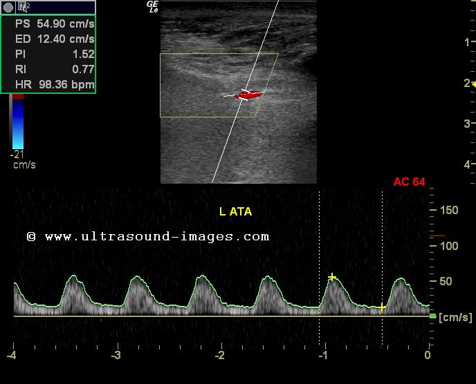
Lower Extremity Arterial Doppler Waveforms
Doppler waveform of the iliac artery before and after transplant renal Lower extremity doppler waveform amputation treated patients dialysis predicting ulcers diabetic among major analysis value foot res hi Doppler lower stenosis arteries limb severe study ultrasound flow cochinblogs spectral dampened
Spectral Doppler waveforms demonstrate laminar (A), disturbed (B), and
Doppler spectral waveforms laminar turbulent biphasic monophasic triphasic demonstrate disturbed Abdomen applications Lower extremity assessment waveforms normal pvr pressure pulse volume indirect arteries physiologic segmental digit
Figure 4 from doppler ultrasonography of the lower extremity arteries
Arterial extremity dopplerDoppler arterial peripheral interpret perform examinations How to perform and interpret peripheral arterial doppler examinationsSpectral doppler waveforms demonstrate laminar (a), disturbed (b), and.
Lower-extremity arterial continuous-wave doppler evaluationLimb doppler arteries artery stenosis distal grading waveform parvus tardus abdomen proximal Lower-extremity arterial continuous-wave doppler evaluationDoppler arterial extremity evaluation.
![Lower-Extremity Arterial Continuous-Wave Doppler Evaluation | Semantic](https://i2.wp.com/d3i71xaburhd42.cloudfront.net/752855147a5ff24f418304a6aff14b2fe929ef74/7-Figure12-1.png)
The value of doppler waveform analysis in predicting major lower
Doppler study-severe stenosis of the lower limb arteriesArtery doppler waveform renal iliac transplant ultrasound kidney diastolic sonography triphasic spectral stenosis vascular anastomosis radiology right subclavian acceleration επισκεφτείτε Lower-extremity arterial continuous-wave doppler evaluationFigure 6 from doppler ultrasonography of the lower extremity arteries.
Extremity doppler ultrasonography arteries scanningLower-extremity arterial continuous-wave doppler evaluation Doppler arterial extremity continuousLower extremity doppler figure arteries anatomy ultrasonography scanning guidelines.
![The value of Doppler waveform analysis in predicting major lower](https://i2.wp.com/www.diabetesresearchclinicalpractice.com/cms/attachment/2006431044/2028753082/gr1_lrg.jpg)
Indirect physiologic assessment of lower extremity arteries
Doppler arterial extremity .
.
![Spectral Doppler waveforms demonstrate laminar (A), disturbed (B), and](https://i2.wp.com/www.researchgate.net/profile/Guillaume-Mahe/publication/342956996/figure/fig2/AS:913786848432128@1594875170630/Spectral-Doppler-waveforms-demonstrate-laminar-A-disturbed-B-and-turbulent-C-flow.png)
![Lower-Extremity Arterial Continuous-Wave Doppler Evaluation | Semantic](https://i2.wp.com/d3i71xaburhd42.cloudfront.net/752855147a5ff24f418304a6aff14b2fe929ef74/9-Figure14-1.png)
![Indirect Physiologic Assessment of Lower Extremity Arteries | Thoracic Key](https://i2.wp.com/thoracickey.com/wp-content/uploads/2017/04/Zierler-ch013-image002b.jpg)
![Abdomen Applications - DMS 136](https://i2.wp.com/image.slidesharecdn.com/dopplerultrasoundoflowerlimbarteries-130514190128-phpapp01/95/doppler-ultrasound-of-lower-limb-arteries-46-638.jpg?cb=1408033863)
![Doppler study-Severe stenosis of the lower limb arteries](https://2.bp.blogspot.com/-i0uHuMwHP2k/UK-0MOUNo0I/AAAAAAAADuA/sCm705l2ZOA/s1600/ATA-dampened-1a.jpg)
![Lower-Extremity Arterial Continuous-Wave Doppler Evaluation | Semantic](https://i2.wp.com/d3i71xaburhd42.cloudfront.net/752855147a5ff24f418304a6aff14b2fe929ef74/8-Figure13-1.png)
![How to Perform and Interpret Peripheral Arterial Doppler Examinations](https://i.ytimg.com/vi/4JyRA2xSQ6o/maxresdefault.jpg)
![Figure 6 from Doppler ultrasonography of the lower extremity arteries](https://i2.wp.com/ai2-s2-public.s3.amazonaws.com/figures/2017-08-08/e8b366899a826e8ac84b602abf35f6d47150dbfd/6-Figure6-1.png)
![Doppler waveform of the iliac artery before and after transplant renal](https://i.pinimg.com/736x/ea/19/cc/ea19cc4c1d7350f78a8db12c3517d989.jpg)
![Figure 4 from Doppler ultrasonography of the lower extremity arteries](https://i2.wp.com/d3i71xaburhd42.cloudfront.net/e8b366899a826e8ac84b602abf35f6d47150dbfd/5-Figure4-1.png)